| #REF | Quantity | Price | |
|---|---|---|---|
| #E-Nov10-1 | 1 mg | € 295.00 | Inquiry |
| #E-Nov10-2 | 2 mg | € 488.00 | Inquiry |
| #E-Nov10-5 | 5 mg | € 1106.00 | Inquiry |
Kit is provided in stable lyophilized form and shipped without dry ice
Bulk quantity available.
You can ask us for a quotation here or write at contact@novocib.com
Download NOVOCIB's Bacterial Luciferase
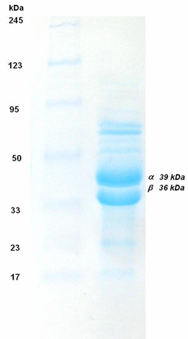
NOVOCIB's bacterial luciferase is purified from a Photobacterium phosphoreum strain isolated from squid by our team and selected for its brightest luminescence. The luxab gene was amplified by PCR and cloned. The sequences of cloned α and β subunits have shown 94% and 92% identity to P24113 and P12744 proteins of Photobacterium phosphoreum (SwissProt Entry).
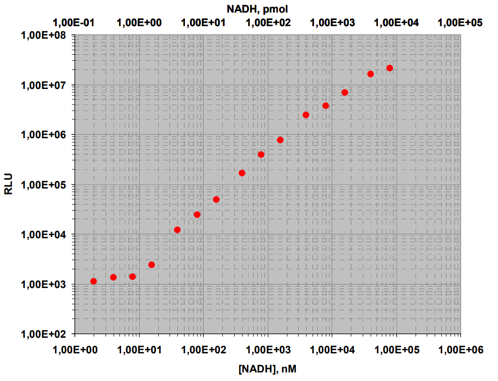
Applications: Applications: In luminescent marine photobacteria, the production of light results from two successive reactions. The first one is catalyzed by the NAD(P)H-FMN oxidoreductase (EC 1.6.8.1), that produces FMNH2 acting as a substrate for the second reaction, which is catalyzed by a luciferase (EC 1.14.14.3) to generate light in the presence of an aliphatic aldehyde and molecular oxygen. In the presence of limiting concentrations of NADH substrate, light intensity is proportional to NAD(P)H concentration. The coupling of bacterial luciferase to FMN-NAD(P)H oxidoreductase has been used to provide ultrasensitive analytical tools for the quantification of NAD(P)H and the substrates of NADH-, NADPH- dependent enzymes (e.g. glucose, lactate, malate, ethanol, sorbitol, oxaloacetate)(1).
Synonyms: Alkanal, reduced-FMN:oxygen oxidoreductase (1-hydroxylating, luminescing)NOVOCIB 's Highly Pure Bacterial Luciferase can be used for NAD(P)H quantification or in dehydrogenase-coupled assays.